Endoscopy
Endoscopy is the insertion of a long, thin tube directly into the body to observe an internal organ or tissue in detail. It can also be used to carry out other tasks, including imaging and minor surgery.
Endoscopies are minimally invasive and involve openings of the body such as the mouth or anus.
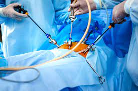
Alternatively, they can be inserted into small incisions, for instance, in the knee or abdomen. Surgery completed through a small incision and assisted with special instruments, such as the endoscope, is called keyhole surgery.
Because modern endoscopy has relatively few risks, delivers detailed images, and is quick to carry out, it has proven incredibly useful in many areas of medicine. Today, an estimated 75 millionTrusted Source endoscopies are carried out each year in the United States.
In this article, we will explain some of the types of endoscopy, why and how they are performed, the general procedure, and any potential risks.
Endoscopies are useful for investigating many systems within the human body; these areas include:
Gastrointestinal tract: esophagus, stomach, and duodenum (esophagogastroduodenoscopy), small intestine (enteroscopy), large intestine/colon (colonoscopy, sigmoidoscopy), bile duct, rectum (rectoscopy), and anus (anoscopy)
Respiratory tract: nose (rhinoscopy), lower respiratory tract (bronchoscopy)
Ear: otoscopy
Urinary tract: cystoscopy
Female reproductive tract (gynoscopy): Cervix (colposcopy), uterus (hysteroscopy), fallopian tubes (falloposcopy)
Through a small incision: abdominal or pelvic cavity (laparoscopy), interior of a joint (arthroscopy), organs of the chest (thoracoscopy and mediastinoscopy)
What is a capsule endoscopy?
Capsule endoscopy was developed in the mid-1990s and involves a wireless camera. The camera is small enough to fit into a capsule (roughly the size of a vitamin tablet) and can, therefore, be swallowed.
As the capsule travels through the digestive tract, it takes thousands of pictures, which are transmitted to a device attached to a wearable belt.
A capsule endoscopy images the small intestine, a difficult region to image using standard endoscopy. It is also very useful for examining the small intestinal mucosa and diagnosing Crohn’s diseaseTrusted Source. The capsule usually passes through the digestive system within 24–48 hours.
While capsule endoscopy has been deemed a sufficient alternative to an esophagogastroduodenoscopy, limitationsTrusted Source are observed, including clear visualization and active movements that need controlling. To remedy this issue, magnetic manipulation is used.